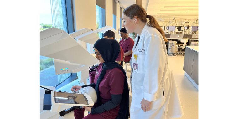
Large, picturesque windows allow natural light to shine on the freshly painted walls of the new Kansas City University College of Dental Medicine (KCU-CDM) on the Farber-McIntire Campus in Joplin, Missouri. The innovative technologies anchored throughout the facility shine in their own right. KCU has revolutionized the dental education experience by providing students with cutting-edge equipment. The dental school ranks among the most technologically advanced educational institutions in the nation.
KCU-CDM boasts four virtual reality (VR) simulators, the most advanced in dental simulation, that are incorporated into the curriculum. Using VR simulators, students can build confidence practicing clinical skills in a learning environment prior to caring for patients in a real-world setting. The technology allows the aspiring dentists to perform multiple repetitions improving their skills prior to providing patient care. Instructors guide students as they progress from basic manual dexterity to restorative procedures to more complex clinical techniques. “They can actually perform on the real anatomy of a person in a virtual reality simulation. It's fantastic because the student can practice the clinical procedure virtually before completing it on a patient,” explained Jacksie Short, DMD, KCU-CDM assistant director of Dental Simulation. Students have practiced restorative procedures in addition to local anesthesia using the VR simulators.
Manikin, or “Charlie,” heads align in rows inside the state-of-the-art dental simulation lab. The Charlie Manikin System compliments the VR technology by replicating a patient’s head and mouth. Typodonts with plastic teeth attach inside each manikin head. These teeth simulate clinical dental conditions such as decay, tartar and periodontal disease. VR simulation gives students the tactical sense of drilling through enamel, dentin and into the pulp chamber.
Intraoral digital scanners with PlanCAD design software allow students to get digital impressions efficiently while eliminating the need for impression trays and messy, uncomfortable impression materials. Images are then used to design crowns and bridges, inlays and onlays, veneers, implants, bite splints and 3D printable models.
Impressions from the scans are sent to the on-site digital milling lab where students learn how to design prosthodontic restorations. Prosthetics can then be created with a 3D printer, producing implant stents, or models for crown preps.
Dental students also have access to a Panceca Viso G7 CBCT/Panoramic/CEPH machine to take a variety of diagnostic images. “Digital imaging plays a key role in today’s oral health care environment,” said Linda Niessen, DMD, CDM founding dean and vice provost of Oral Health Affairs. “The investment that KCU has made in this advanced dental technology is providing an outstanding educational opportunity for our students. It is improving the efficiency of patient care, and enabling the College of Dental Medicine to become a center of excellence in the region improving oral health and increasing access to oral health care.”


(0) Comments